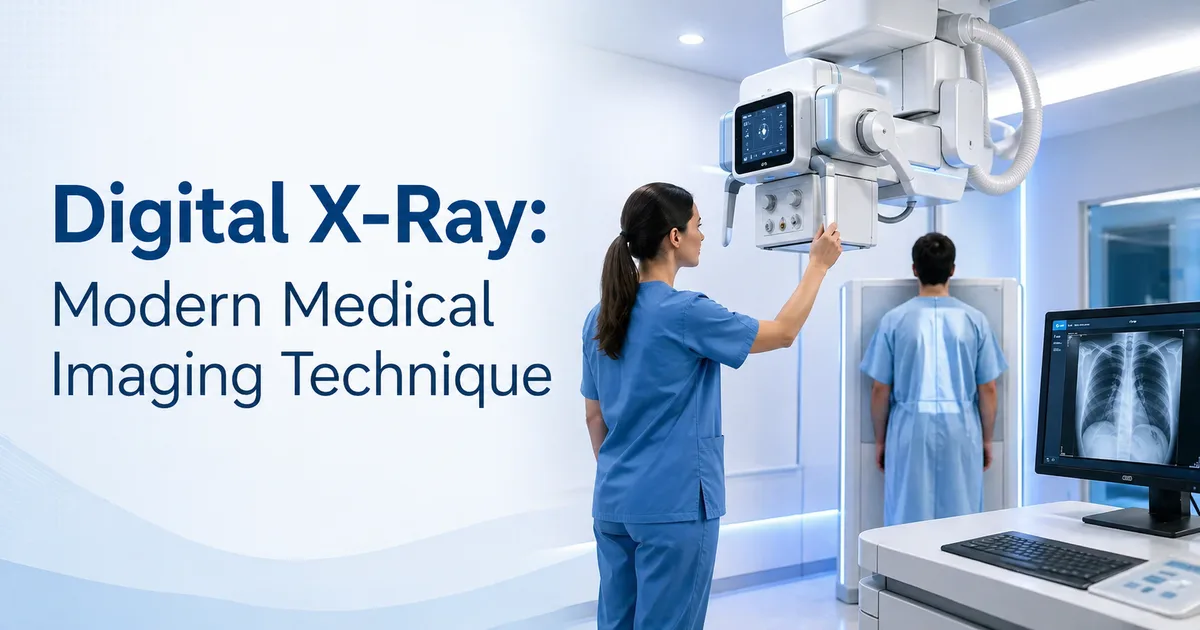
What Is Digital X-Ray? How Does It Work?
Digital X-ray is a form of radiography that captures X-ray images electronically, storing them as digital files instead of on photographic film. This technology allows doctors to view, enhance, and analyse images on computer screens instantly.
How Digital X-Ray Works:
- An X-ray beam passes through the body part being examined
- A digital detector (not film) captures the radiation
- The detector converts X-ray energy into electronic signals
- A computer processes the signals and creates a digital image
- Images can be viewed instantly on screens and archived digitally
Digital X-Ray vs. Traditional X-Ray: Key Benefits
|
Feature
|
Digital X-Ray
|
Traditional X-Ray
|
|
Image Quality
|
Superior detail and clarity
|
Good but limited detail
|
|
Radiation Dose
|
50-80% lower radiation exposure
|
Higher radiation dose
|
|
Speed
|
Instant image availability
|
24-48 hours for film development
|
|
Storage
|
Digital files (compact, searchable)
|
Physical film (bulky, deteriorates)
|
|
Image Enhancement
|
Brightness, contrast, zoom adjustable
|
Limited adjustment options
|
|
Cost
|
Lower long-term cost per image
|
Higher per-image cost
|
|
Patient Safety
|
Minimal radiation, safer
|
More radiation exposure
|
|
Doctor Efficiency
|
View instantly from anywhere
|
Must wait for film processing
|
|
Comparison
|
Easy to compare past images
|
Difficult to locate old films
|
|
Environmental
|
No chemical processing
|
Chemical waste from processing
|
Medical Uses of Digital X-Ray
Digital X-ray is used across many medical specialities for diagnosis and monitoring:
1. Cancer Diagnosis & Staging
- Chest X-ray: Detect lung cancer, metastasis to lungs
- Bone X-rays: Identify bone metastasis in cancer patients
- Abdominal X-rays: Assess GI and abdominal tumours
2. Fracture & Trauma Diagnosis
- Detect broken bones and joint injuries
- Monitor healing progress
3. Infection & Inflammation
- Pneumonia and respiratory infections
- Chronic disease monitoring
4. Pre-Operative Planning
- Assess surgical anatomy before oncology procedures
- Guide radiation therapy planning
Is Digital X-Ray Safe? Radiation Exposure
Yes, digital X-ray is safe when used appropriately. Here’s what you need to know about radiation exposure:
Radiation Dose Comparison:
Single chest X-ray: ~0.1 millisieverts (mSv) — equivalent to 10 days of natural background radiation
Annual background radiation: ~2.4 mSv — dose everyone receives from nature
CT scan: ~7 mSv — 70 times higher than chest X-ray
Digital X-rays use 50-80% less radiation than traditional film X-rays while producing superior images.
Safety Precautions:
- Lead aprons protect sensitive organs
- Pregnant patients should inform staff (though risk is low for single X-rays)
- Technicians are trained to minimise unnecessary exposure
- Multiple X-rays should be justified by medical need
Digital X-Ray at Onco-Life Cancer Centre
Onco-Life Cancer Centre is equipped with state-of-the-art digital radiography systems as part of our comprehensive diagnostic imaging capabilities.
Our Digital X-Ray Services:
- Instant image availability: Results available to doctors within minutes
- Expert interpretation: Analysed by experienced oncology radiologists
- Integration with other imaging: Coordinated with PET-CT, MRI, ultrasound
- Radiation-optimised protocols: Minimise exposure while maximising diagnostic information
- Digital archiving: Secure storage and easy retrieval of past studies
Digital X-ray is often the first imaging modality when a patient presents with symptoms suggestive of cancer or when monitoring treatment response.
Digital X-Ray vs. Other Imaging Modalities
When Is Digital X-Ray the Best Choice?
- First-line imaging: Quick, low-dose screening when initial assessment needed
- Lung cancers: Detect masses and pleural involvement
- Bone metastasis: Identify skeletal involvement in disseminated cancer
- Post-operative monitoring: Track complications after surgery
When Other Imaging Is Preferred:
- Soft-tissue detail: MRI provides superior soft tissue contrast
- Metabolic information: PET-CT shows functional activity
- Fast screening: Ultrasound faster and less expensive
NEED DIAGNOSTIC IMAGING FOR CANCER EVALUATION?
Onco-Life provides comprehensive diagnostic imaging including digital X-ray, PET-CT, MRI, and ultrasound with expert interpretation by our oncology radiologists.
Schedule your imaging appointment at Onco-Life Cancer Centre — Talegaon, Satara, Chiplun, or Wagholi
About Onco-Life Cancer Centre
Onco-Life Cancer Centre is Maharashtra’s largest and most trusted dedicated oncology hospital network with three National Accreditation Board for Hospitals and Healthcare Providers (NABH) accreditations. We are members of the National Cancer Grid (NCG) and have treated over 1,20,000 patients.
We offer comprehensive diagnostic imaging services including the digital radiography, PET-CT (Positron Emission Tomography), MRI, ultrasound, and digital pathology across four centres in Maharashtra. Our imaging services support accurate diagnosis and treatment planning for all cancer types.
DISCLAIMER
This article is for informational purposes only and is not a substitute for professional medical advice, diagnosis, or treatment. Always seek the guidance of a qualified healthcare provider with any questions regarding a medical condition.